What Surrounds the Individual Muscle Cell
T-tubules in cardiac muscle are bigger and wider than those in skeletal muscle but fewer in number. The nuclei myonuclei of a multinucleated muscle fiber are no longer.

Skeletal Muscle Tissue
This organelle surrounds the myofibril like a loosely knit sweater surrounds your arm.

. And are open at the cell surface to the extracellular fluid that surrounds the cell. And endomysium surrounding muscle fibers. The development of a muscle fiber is not complete however with the peripheral migration of the nuclei of the myotube.
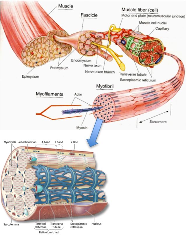
The cell that surrounds normal breast ducts and and lobules. Connective tissue which surrounds the muscle at many levels is organized into epimysium surrounding the whole muscle. Skeletal muscles have an abundant supply of blood vessels and nerves.
In the centre of the cell they join running into and along the cell as a transverse-axial network. Japanese honey bees swarm hornets killing them with heat. Myoepithelial cells are still present around ducts that contain carcinoma in situ however invasive carcinomas lack myoepithelial cells.
Within the fasciculus each individual muscle cell called a muscle fiber is surrounded by connective tissue called the endomysium. From the cell lineage studies of Conklin and others see Chapter 1 it was known that only one pair of blastomeres the posterior vegetal pair B41 in the 8-cell tunicate embryo is capable of producing tail muscle tissue. Masato Ono Tamagawa University Rhodopsin is known to be involved in temperature sensing in fruit flies and the authors speculate it may function.
In the embryo a skeletal muscle fiber begins as a single cell called a myoblastIndividual myoblasts begin to fuse with one another forming elongated cells called myotubes in which dozens of nuclei are lined up in a central row Fig. At each increasing size scale extracellular matrix ECM the surrounding connective tissue encapsulates muscle structures. Myoepithelial cells have some contractile properties and also produce the normal basement membrane that surrounds the ducts and lobules.
Skeletal muscle cells occur in the form of multinucleated fibers that can be up to several centimeters long. Within the fasciculus each individual muscle cell called a muscle fiber is surrounded by connective tissue called the endomysium. It forms an internal compartment the.
Generally an artery and at least one vein accompany each nerve that penetrates the. As discussed in Chapter 1 the B41 blastomere pair contains the yellow crescent cytoplasm that correlates with muscle determination When Whittaker. The final stage in the differentiation of the skeletal muscle cell.
Before a skeletal muscle fiber can contract it has to receive an impulse from a neuron. Each compartment contains a bundle of muscle fibers. Individual cardiac muscle cells are joined at their ends by intercalated discs to form long fibers.
Each bundle of muscle fiber is called a fasciculus and is surrounded by a layer of connective tissue called the perimysium. A recent study suggests thermosensing genes are key to the balling behavior. The DMD gene regulates encodes for the production of dystrophin a protein that appears to play an essential role in maintaining the integrity of cell membrane in skeletal voluntary and cardiac muscle cells.
Dystrophin is found attached to the inner side of.
How Muscles Work Part 1 Of 2 Shapelog

10 2 Skeletal Muscle Anatomy Physiology

Skeletal Muscle Anatomy And Physiology I Archived


Comments
Post a Comment